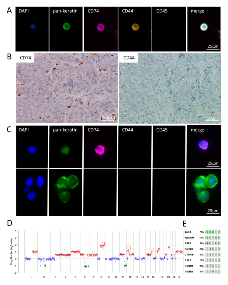
Figure 3.
Matched samples of blood CTCs, BM and CSF-CTCs of breast cancer patient P6. (A) Exemplary CTC positive for keratins, CD74 as well as CD44 and negative for CD45 (leukocyte exclusion marker). (B) Matched BM tissue shows no expression of CD74 and CD44. (C) Matched liquor samples show single CTCs as well as clusters. Only one liquor DTC was positive for CD74, whereas all DTCs were negative for CD44 and CD45. (D) Copy number alteration profile of a single CTC of P6 showing numerous large chromosomal losses and gains, including a high-level amplification of chromosomes 8 (MYC loci) and 12 and 17 (HER2) seen in all CTCs. (E) Most common identified mutation in 10 CTCs analyzed (green: missense mutation (unknown significance); dark green: missense mutation (putative driver); grey: truncating mutation (unknown significance).

