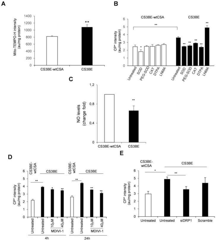
Figure 4.
(A) Mitochondrial ROS levels assessed by EPR technique by using the cyclic hydroxylamine spin probe Mito-TEMPO-H in CS3BE and CS3BE-wtCSA cells. (B) ROS species characterization in CS3BE and CS3BE-wtCSA cells by competition with the suitable scavengers for O2-• and H2O2 as indicated. SOD, superoxide dismutase; PEG-SOD, superoxide dismutase conjugated to polyethylene glycol (PEG); CAT, catalase; DTPA, diethylenetriamine pentaacetic acid, and NMA, N-monomethyl-L-arginine. (C) NO levels in CS3BE and CS3BE-wtCSA cells measured by DAF-FM-DA. D, E. ROS levels assessed by EPR technique by measuring the intensity of the formed nitroxide 3-carboxyproxyl radical in CS3BE and CS3BE-wtCSA cells after treatment with MDIVI-1 (D) or DRP1 silencing (E). The reported values are mean ± SD of three independent experiments; * p < 0.05, ** p < 0.01 by paired Student t-test.

