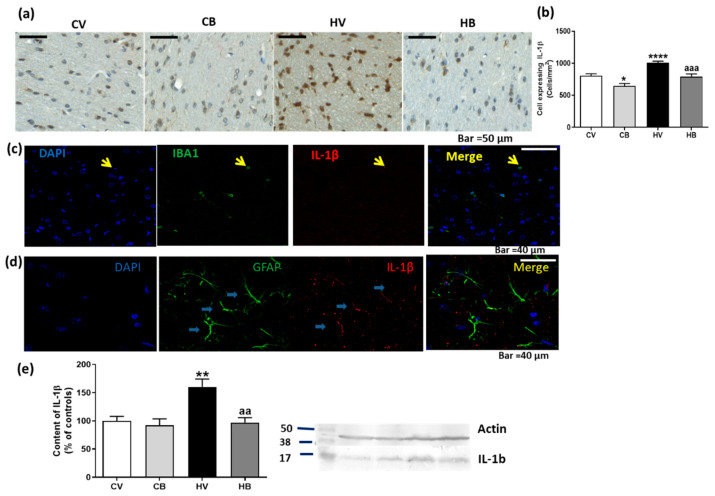
Figure 4.
Effect of bicuculline on the content of IL1β in white matter of the cerebellum. Immunohistochemistry was performed using antibody against IL-1β. Representative images of IL-1β staining in white matter of the cerebellum are shown (a). The number of cells expressing IL-1β was quantified. Data are the mean ± SEM of 3–4 rats per group (b). Double immunofluorescence of IL-1β and Iba1 is shown in (c) and of IL-1β and GFAP in (d), confirming the expression of IL-1β in both microglia and astrocytes. The content of IL-1β was also analyzed by Western blot in the total cerebellum. Values are mean ± SEM of 12–14 samples per group (e). Values significantly different from control rats are indicated by asterisks: * p < 0.05, ** p < 0.01, **** p < 0.0001. Values significantly different from hyperammonemic rats are indicated by “aa”, p < 0.01, “aaa”, p < 0.001. CV, control vehicle; CB, control treated with bicuculline; HV, hyperammonemic rats with vehicle; HB, hyperammonemic rats treated with bicuculline.

