Figure 2.
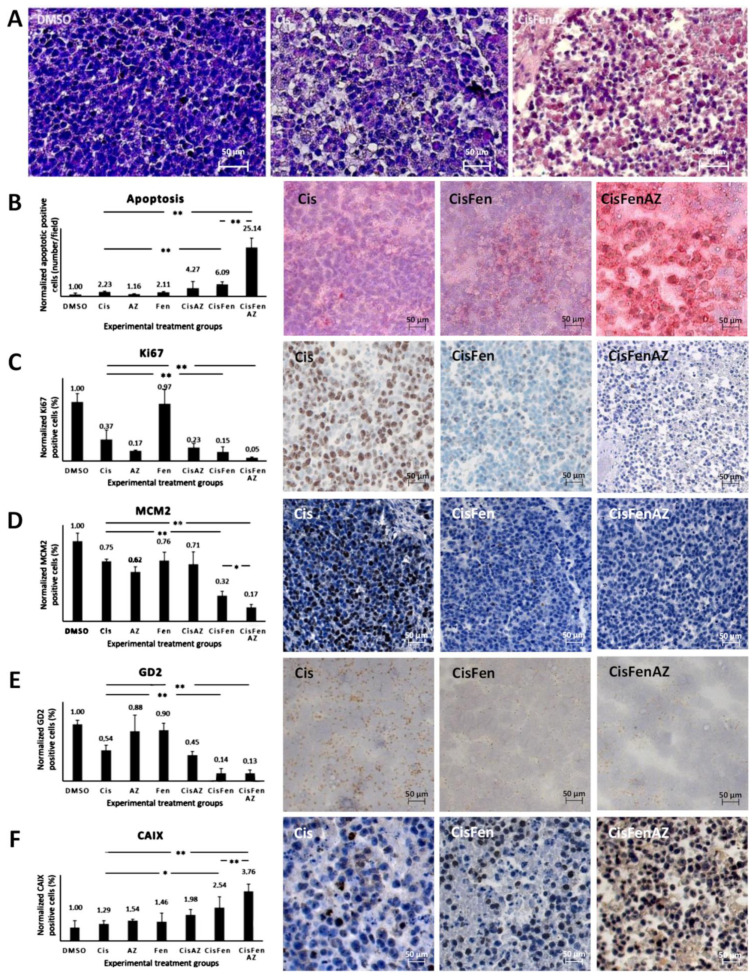
Hystochemical analysis of SKNBE2 nodules. Representative images of hematoxylin staining of SKNBE2 nodules (A) from vehicle-(DMSO), cisplatin-(Cis) and cisplatin, fendiline, and acetazolamide-(CisFenAZ) treated mice. The percentage of pycnotic nuclei and mass disaggregation is strongly increased by CisFenAZ treatment. Quantification of apoptotic cells by TUNEL assay in SKNBE2 nodules after treatments (B). Values refer to the amount of TUNEL-positive cells per field (left plot). Panels on the right show representative images of cisplatin-(Cis), cisplatin/fendiline-(CisFen) and cisplatin/fendiline/acetazolamide-(CisFenAZ) treated mice. The number of apoptotic cells in cisplatin-treated tumors was increased by the addition of either acetazolamide (4.27 vs. 2.23) or fendiline (6.09 vs. 2.23); remarkably, the contemporary administration of acetazolamide and fendiline with cisplatin produced a strong increase of apoptotic cells (25.14 vs. 2.23). Analysis of Ki-67 (C), MCM2 (D), GD2 (E), and CAIX (F) expression in SKNBE2 nodules. Plots on the left quantify the amount of positive cells per field. Panels on the right show representative images of cisplatin-(Cis), cisplatin/fendiline-(Cis/Fen) and cisplatin/fendiline/acetazolamide-(CisFenAZ) treated mice. Cisplatin produced a significant reduction of Ki-67, MCM2, and GD2. The expression of Ki-67 was strongly reduced by acetazolamide alone and almost abolished when acetazolamide was added along with fendiline and cisplatin (C). MCM2 reduction by cisplatin was strengthened by fendiline and strengthened further by fendiline/acetazolamide (D). The effect of cisplatin on reducing GD2 was strengthened by fendiline only (E). All treatments except DMSO produced a significant increase in CAIX expression; the addition of acetazolamide induced a further highly significant CAIX increase, compared with CisFen (* p < 0.05, ** p < 0.001). Space bar—50 µM.

