Figure 5.
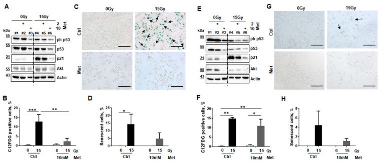
Metformin treatment prevents RT-induced senescence in normal epithelial lung cell cultures. The impact of metformin treatment for RT-induced senescence formation was investigated in cultures of lung epithelial cell lines derived from normal lung epithelium (BEAS-2B and HSAEC1-KT) by analyzing protein expression levels of senescence markers via Western blot (A,E), C12FDG staining and flow cytometry (B,F), and SA-betagal activity staining (C,D,G,H). p21 and p53 expression levels and respective levels of the cell survival marker AKT/PKB were analyzed by Western Blots at 96 h post RT and treatment in BEAS-2B (A) and HSAEC1-KT (E) cells. Representative blots of 3 independent experiments are shown. Dashed lines indicate different areas of the same blot. Radiation-induced senescence formation was analyzed by C12FDG treatment prior flow cytometric analyses in BEAS-2B (B) and HSAEC1-KT (F), eight days post treatment. Data showed mean values ±SEM of six independent experiments. p-values indicate: * p ≤ 0.05, ** p ≤ 0.01, *** p ≤ 0.001 by two-way ANOVA with post hoc Tukey’s multiple comparison test. Following SA-betagal activity staining, senescent BEAS-2B (C) and HSAEC1-KT (G) cells were visualized in in blue. Representative pictures are shown (scale bar: 200 µm). (D,H) Senescence formation was quantified by counting the numbers of SA-betagal-positive and -negative epithelial cells. Data showed mean values ±SEM of 3–5 independent experiments. p-value indicates: * p ≤ 0.05 by two-way ANOVA with Tukey’s multiple comparison test.

