Figure 1.
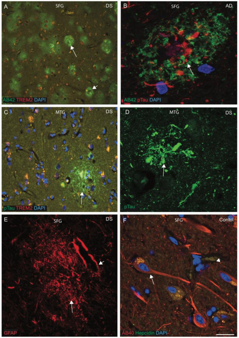
Characterisation of senile plaques (SP) in DS brain. To characterise the SP formation, the DS, AD and control brain sections from the superior frontal gyrus (SFG) and mid temporal gyrus (MTG) were labelled with double immunofluorescence using anti-Aβ42, phospho-tau (with AT8 antibody), anti-hepcidin and anti-TREM2, then counterstained with DAPI for nuclei (blue) and imaged with confocal microscopy. In DS brain, sections were stained with Aβ42 antibody, and an abundant cotton wool appearance of senile plaques (SP) was seen throughout the cortex (A, white arrows highlight the selected area of plaque formation). One AD brain section when stained with Aβ42 and pTau and analysed by confocal microscope showed a mature SP that contained many pTau-positive dystrophic neurites in the centre, with distinct Aβ42-positive astrocytes in the surrounding area (B). Another DS brain section from MTG was stained with pTau and TREM2, and neurofibrillary tangle (NFT) was visible in the core of the plaques, with some co-localisation with TREM2 in the SP and close by (C). In a confocal image of DS, MTG showed pTau-positive NFT visible very close to blood vessels (D). SFG samples from DS brain when stained with GFAP showed astrocytes surrounding the endothelial layer of blood vessels (E, short arrow highlights the blood vessel wall). One SFG from a normal brain section was stained with Aβ40 and hepcidin, and amyloid proteins were found to be evenly distributed in the neuronal cell body, axons and dendrites, whereas hepcidin was present in the cell bodies (F). Scale bar: (A,C) = 50 μm, (B–F) = 25 μm.

