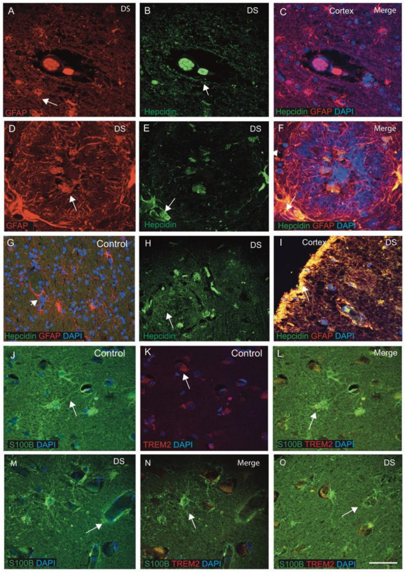
Figure 2.
Hepcidin and S100β-positive activated damaged astrocytes seen around the wall of blood vessels in DS. DS and control brain sections from the cortex and close to the blood vessels labelled with double immunofluorescence (IF) using anti-hepcidin (green), anti-GFAP (red), counterstained with DAPI for nuclei (blue) and imaged with confocal microscopy. In DS brain sections, GFAP-positive activated astrocytes were present around the SP and surrounding the blood vessels (A–C, indicated with an arrow). Hepcidin expression was visible in the endothelial cells of blood vessels (B) and co-localised with the GFAP-positive astrocytes in the DS (E,F). In the senile plaques, GFAP-positive astrocytes were well-organised around the plaque formation (D–F). In the control brain section, normal astrocytes with fine processes were visible and co-localised with hepcidin in the cell bodies (G). A large number of small vesicles carrying hepcidin were present in the SP of the DS cortex and its surroundings (H). Very strong co-localisation of hepcidin with GFAP was noticed in layers I and II of the cortex (I). DS and control brain sections from SFG were stained with S100β and inflammatory protein TREM2. In control brain sections, thin filamentous processes of end-feet of normal astrocytes were present surrounding the blood vessels, positive for S100β and directly connected to the endothelial lining of blood vessels (J). TREM2 was present in the end-feet of astrocytes carrying soluble TREM2 (K) and co-localised in the cell bodies (J–L). In a DS brain section, S100β-positive activated astrocytes were found disorganised and aggregated around the blood vessel walls, indicating severe damage of endothelial cells of blood vessels (M–O). Scale bar: (A–C) = 25 μm, (D–F) = 15 μm, (G–O) = 30 μm.

