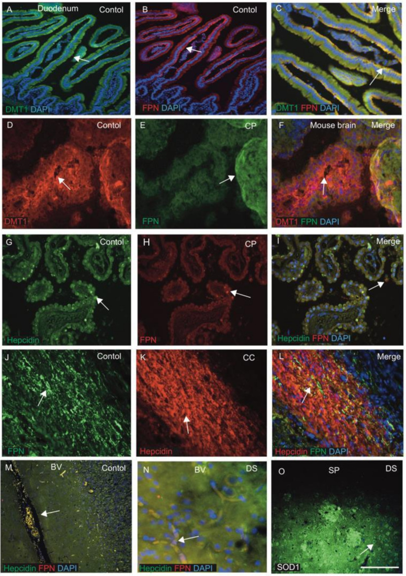
Figure 4.
DMT1, FPN and hepcidin regulate endothelial transport and SOD1 modulates redox balance in DS brain. Tissue sections from mouse duodenum and choroid plexus (CP) were stained by IF using anti-DMT1 and anti-FPN antibodies and counterstained with DAPI for nuclei (blue). In duodenum, DMT1 staining was seen in the columnar cells within the crypts, particularly in the apical surface (A) and FPN at the basolateral site and in the central core of lamina propria containing blood vessels (B,C). The choroid plexus (CP) sections from mouse brain (D–F) stained with DMT1 were expressed in the monolayer of the epithelial cells of CP on luminal sites, whereas FPN was found in the abluminal site of CP, with some co-localisation observed (D–F). Similarly, for human CP when stained with hepcidin and FPN antibodies, hepcidin was visible in the epithelial cells of CP and FPN was seen in the macrophages very close to CP (G–I). Hepcidin and FPN are both visible in the oligodendrocytes located in the corpus callosum (CC), suggesting that iron transport may take place through CC (J–L). Hepcidin and FPN were visible in the blood vessels (M,N). A brain section from the cortex close to a SP was stained with SOD1 antibody, and very strong SOD1protein was seen in the plaque and in the peripheral neurons (O). Scale bar: (A–F) = 20 μm, (G–L) = 25 μm, (M) = 50 μm, (N,O) = 20 μm.

