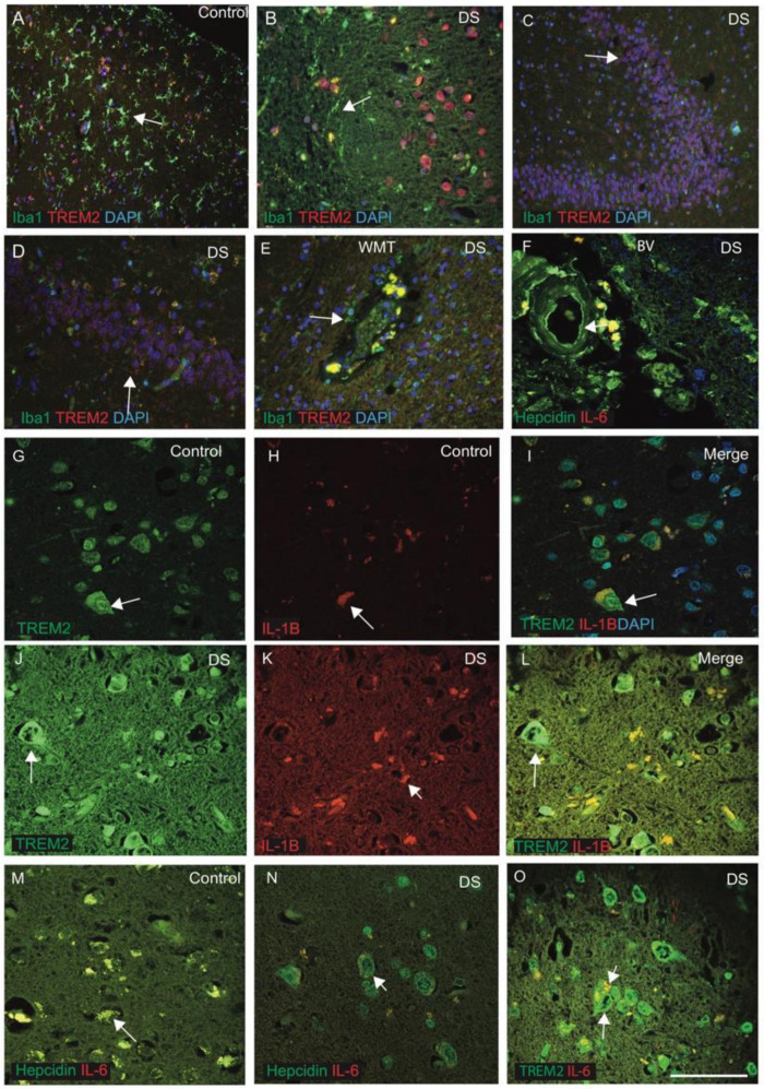
Figure 5.
Inflammatory changes in the DS brain were identified by TREM2, IL-1β and IL-6. To investigate the inflammatory changes in DS, the brain sections from DS and control subjects were stained with microglia marker (Iba1) and TREM2. In control brains, Iba1-positive ramified microglia were visible in the cortex, whereas TREM2 was noticed in the neurons close to the blood vessels (A). In the DS brain sections, activated microglia was visible around the SP and limited co-localisation with TREM2 in the microglia (B). For sections from the HP and DG of DS brain when stained with Iba1 and TREM2, Iba1-positive activated microglia were present close to the neurons, whereas TREM2 expression was visible only in the damaged DG granule cells with limited co-localisation (C,D). The white matter (WM) close to the blood vessels (BV) in the DS brain showed large numbers of Iba1-positive microglia, close to the blood vessels, that might be involved in the clearing process of damaged oligodendrocytes, and TREM2-positive macrophages might be entering from blood vessels (E). For another DS brain section, close to the BV, when stained with hepcidin and IL-6, the staining patterns suggested that both proteins might be entering from the blood vessels (F). The brain sections from controls and DS subjects (from cortex) were stained with TREM2 and pro-inflammatory marker IL-1β. In control, TREM2 was visible in the neurons (G) and in other glial cells, and only very faint IL-1β expression was seen (H), with limited co-localisation in the cell bodies (I). In DS brain, TREM2 expression was seen in the neurons, whereas IL-1β was noticed in the microglia, close to the blood vessels and with some co-localisation noted (J–L). Similarly, for control and DS brain sections, when stained with hepcidin and IL-6, both the proteins were present in the cells close to the blood vessels and with some co-localisation (M), whereas in the DS brain, damaged neurons with a halo around the nucleus were positive for hepcidin (N), and IL-6 was visible in the microglia (N,O). Scale bar: (A–C) = 50 μm, (D–F) = 30 μm, (G–O) = 20 μm.

