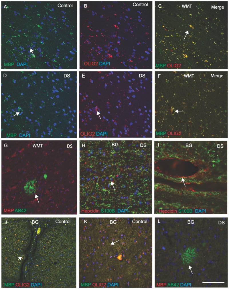
Figure 6.
OLIG2 defect impairs myelination in DS brain. For DS and control white matter (WM) in the cortex and in the basal ganglia (BG), OLIG2 and myelin basic protein (MBP) expression were analysed by IF and counterstained with DAPI for nuclei (blue). In the control brain, MBP expression was seen in the myelinated sheath of oligodendrocytes, with OLIG2 localising in the cell bodies (A–C), and in the DS brain, myelin sheaths appeared to be shrunken and damaged (D–F). In DS brain, Aβ42-positive SPs were visible in the WM of the cortex (G). For another DS brain section from BG, when stained with hepcidin and S100β, both proteins were seen in the WM, and in particular, S100β was present in the oligodendrocytes (I), in astrocytes (H,I), whereas hepcidin was seen in the blood vessels walls (H,I). In control brain, particularly in the BG, both proteins, MBP and OLIG2, were seen in WMT (J,K), whereas, in DS brain, Aβ42-positive SPs were visible (L). Scale bar: (A–F) = 20 μm, (G–L) = 30 μm.

