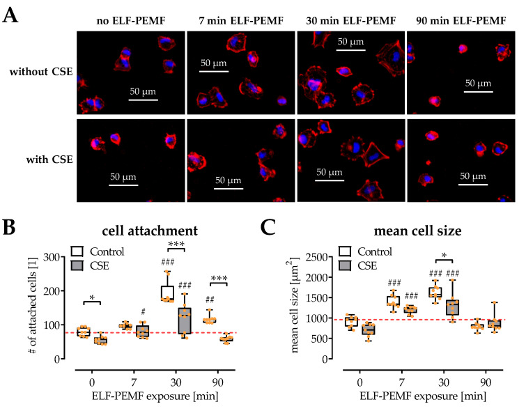
Figure 2.
Influences of 16 Hz ELF-PEMFs (0, 7, 30, and 90 min) and 5% cigarette smoke extract (CSE) on SCP-1 cell adhesion and spreading after 4 h. (A) Representative images (400× magnification) of the fluorescence staining for cytoskeleton (phalloidin-TRITC, 2 μg/mL, red) and nuclei (Hoechst 33342, 2 μg/mL, blue). (B) Automated quantification of adherent nuclei, and (C) the mean size of attached SCP-1 cells using the ImageJ software. N = 3, n = 3. Data are presented as box plots (Min to Max with single data points). Data were compared by non-parametric two-way ANOVA followed by Tukey’s multiple comparison test: * p < 0.05 and *** p < 0.001 as indicated; # p < 0.05, ## p < 0.01, and ### p < 0.001 as compared to the respective control (no ELF-PEMF).

