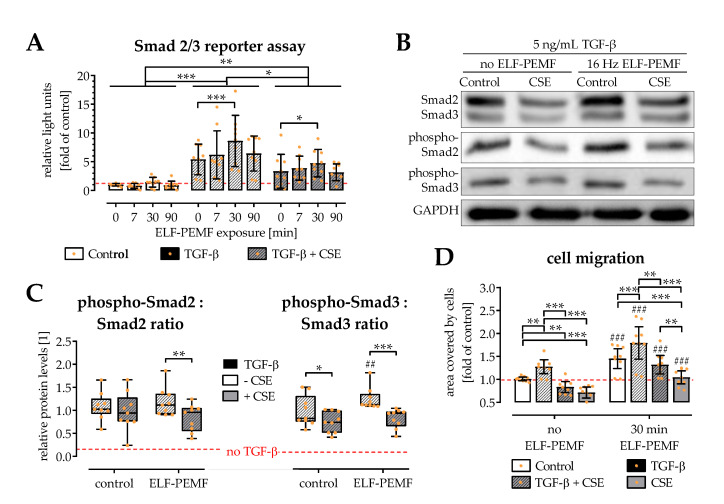
Figure 4.
Canonical (Smad2/3) TGF-β signaling affected by 5% cigarette smoke extract (CSE) is fortified by exposure to the 16 Hz ELF-PEMFs. (A) An adenoviral reporter assay was used to quantify canonical (Smad2/3) TGF-β signaling in SCP-1 cells exposed to 5 ng/mL TGF-β, 5% CSE, and/or the 16 Hz ELF-PEMFs (0, 7, 30, 90 min daily exposure) for 72 h. Western blot was used to confirm phosphorylation of Smad2 and Smad3 in the cells with 30 min daily exposure to the 16 Hz ELF-PEMF. (B) Representative image of the Western blot. (C) Signal intensities were quantified with the ImageJ software and the ratio of phosphorylated-Smad2 to Smad2 and phosphorylated-Smad3 to Smad3 were determined. (D) SCP-1 cell migration was determined using the cell migration assay kit. Cells invading the migration zone were quantified using the ImageJ software. N = 3, n = 3. Data are presented as box plots (Min to Max with single data points). Data were compared by non-parametric two-way ANOVA followed by Tukey’s multiple comparison test: * p < 0.05, ** p < 0.01, and *** p < 0.001 as indicated; ## p < 0.01 and ### p < 0.001 marking the ELF-PEMF effect.

