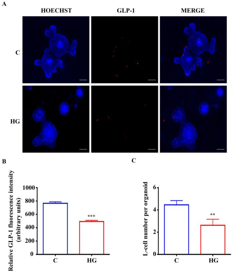
Figure 6.
Expression of GLP-1 in small intestinal organoids. (A) Representative images of controls and organoids treated with high glucose (35 mM) for 48 h. L-cells, inside organoids, are labeled in red by GLP-1 expression. Nuclei are labeled by Hoechst. Scale bar, 50 μm. (B) Quantification of the GLP-1 fluorescence intensity. (C) L-cell numbers in control organoids and organoids treated with high glucose. (n = 6). Data are expressed as means ± SEM. Unpaired Student t-test: ** p < 0.01, *** p < 0.001. C = Control (17.5 mM glucose). HG = High Glucose (35 mM glucose).

