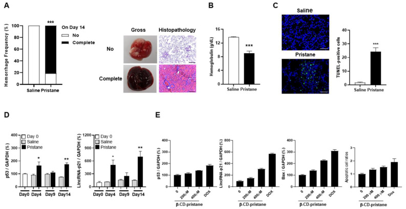
Figure 3.
LincRNA-p21 expression in pristane-induced model of DAH and hydrophilic pristane-stimulated alveolar epithelial cells. (A) Hemorrhage frequencies in PBS- and pristane-injected C57BL/6 mice on day 14 (left panel). Representative gross and histopathology findings in the lungs with no or complete hemorrhage (right panel). (B) Hb levels of PBS- and pristane-injected C57BL/6 mice on day 14. (C) Representative TUNNEL staining for apoptotic cells in lung tissues from PBS- and pristane-injected mice (left panel, ×400). Bars shown on photomicrographs corresponding to 20 µm. Numbers of TUNEL-positive cells (right panel), as determined by averaging the number from 3 fields (×400) of the highest density of positively stained cells in each section. (D) Serial p53 (left panel) and lincRNA-p21 (right panel) pulmonary expression levels on day 0, 4, 9 and 14 from PBS- and pristane-injected mice. (E) p53, lincRNA-p21 and Bax expression as well as apoptotic cell ratios in alveolar epithelial cells stimulated with different concentrations of hydrophilic pristane for 24 h. Relative abundance of a measured gene expression was normalized by GAPDH gene from each sample. The average levels of mouse lung tissues on day 0 and expression levels of alveolar epithelial cells without stimulation were determined as 100%. Values are mean ± SEM. Sixteen mice per group in (A,B), and 5 mice per group in (C,D). All of the in-vivo results in (A–D) and in-vitro results in (E) in Figure 3 were representative of two and three independent experiments, respectively, with similar findings. * p < 0.05, ** p < 0.01, *** p < 0.001.

