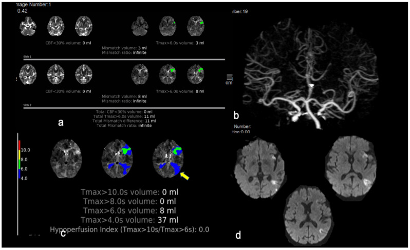
Figure 2.
This is an illustrative case of a patient fulfilling both neuroimaging and clinical EXTEND eligibility criteria who was treated successfully with intravenous thrombolysis in the extended time window. An 80-year-old woman was transferred from an island to the emergency department 5 h after an acute onset of expressive aphasia, mild right facial paresis, and mild right upper arm paresis (ΝΙHSS score 9 points). (a) Her CT-perfusion mismatch map post-processed with RAPID software demonstrated a hypoperfused region of 11 mL in the Broca’s area (shown in green) and no area of reduced cerebral blood flow, resulting in a 11 mL mismatch difference (infinite mismatch ratio). (b,c) CT angiogram revealed no large vessel occlusion. The patient fulfilled all EXTEND eligibility criteria; IVT with alteplase started 5 h and 45 min after symptom onset with partial resolution of symptoms at the end of tPA infusion (NIHSS-score of 6 points). (d) Repeat MRI at 24 h demonstrated a small insular infarct and another acute infarct in the left temporoparietal region which was captured in the Tmax maps of initial perfusion imaging as Tmax > 4 s prolongation (c/arrow). The patient’s mRS-score at three months was 0.

