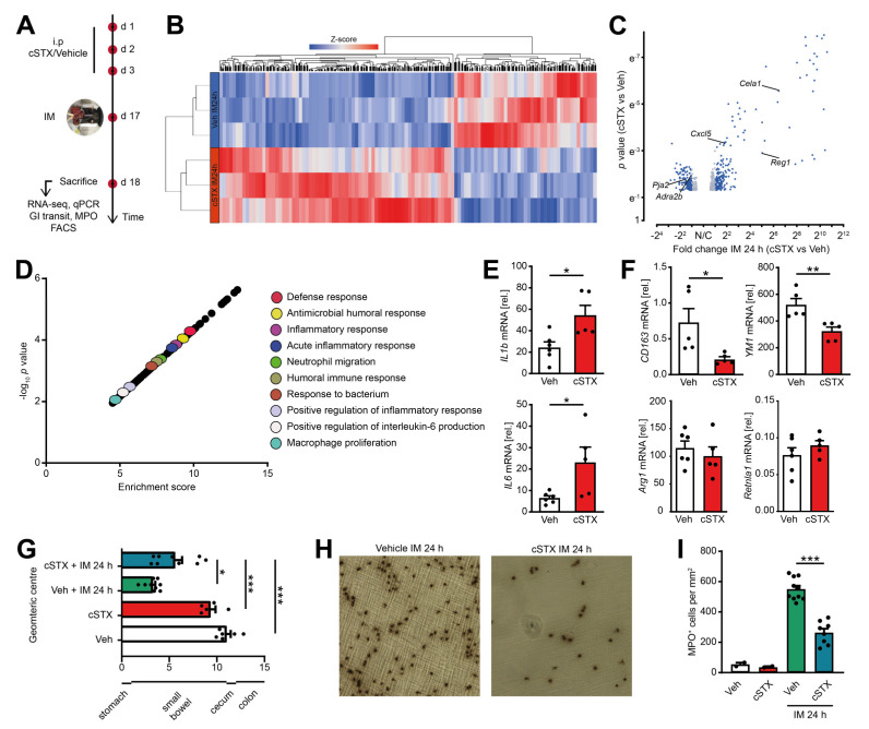
Figure 5.
Effects of sympathetic neuronal depletion in the effector phase of POI. Mice were subjected to cSTX or vehicle (Veh) treatment before they underwent IM. (A) The experimental scheme to analyze the role of sympathetic innervation in POI. (B) Heat map analysis of differentially expressed genes showed the hierarchical clustering between vehicle- and cSTX-treated mice 24 h after IM. (C) Volcano plot of 456 differentially expressed genes between cSTX and vehicle-treated mice 24 h after IM, showing more than half of the genes upregulated in the cSTX-treated mice 24 h after IM. (D) Functional enrichment of immune-related GO terms. (E,F) qPCR analysis showing the mRNA expression of pro-inflammatory (E) and anti-inflammatory markers (F) from the small-intestinal muscularis samples of vehicle- (n = 6) (white) or cSTX-treated (n = 5) (red) mice 24 h after IM. Values in each column are shown as mean ± SEM, and statistical analysis was carried out by unpaired t-test (*p < 0.05, **p < 0.01, and ***p < 0.001) (E,F) comparing vehicle-treated mice without IM versus indicated groups. (G) GI transit time calculated 24 h after IM and shown as the geometric center of distribution of FITC dextran in the stomach (st), small intestine, cecum (c), and colon (n = 9). (H) Ileal muscularis whole mounts of vehicle- (left) and cSTX-treated (right) mice 24 h after IM showing myeloperoxidase-staining (MPO) for polymorphonuclear neutrophils (PMNs). (I) Quantification of MPO+ leukocytes in cSTX compared to vehicle-treated mice 24 h after IM. The graphs are plotted as mean ± SEM, and statistical analysis was carried out by two-way ANOVA (* p < 0.05, ** p < 0.01, and *** p < 0.001), (G,I) comparing vehicle-treated mice with indicated groups.

