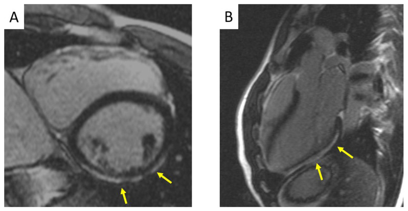
Figure 1.
Cardiac magnetic resonance in a patient with a desmosomal gene related biventricular ACM showing the typical LV LGE pattern. (A) Short axis view demonstrating subepicardial LGE at the LV mid-inferolateral segments. (B) Three-chamber view exhibiting extensive LGE stria at the LV posterolateral wall. ACM = arrhythmogenic cardiomyopathy; LGE = late gadolinium enhancement; LV = left ventricle.

