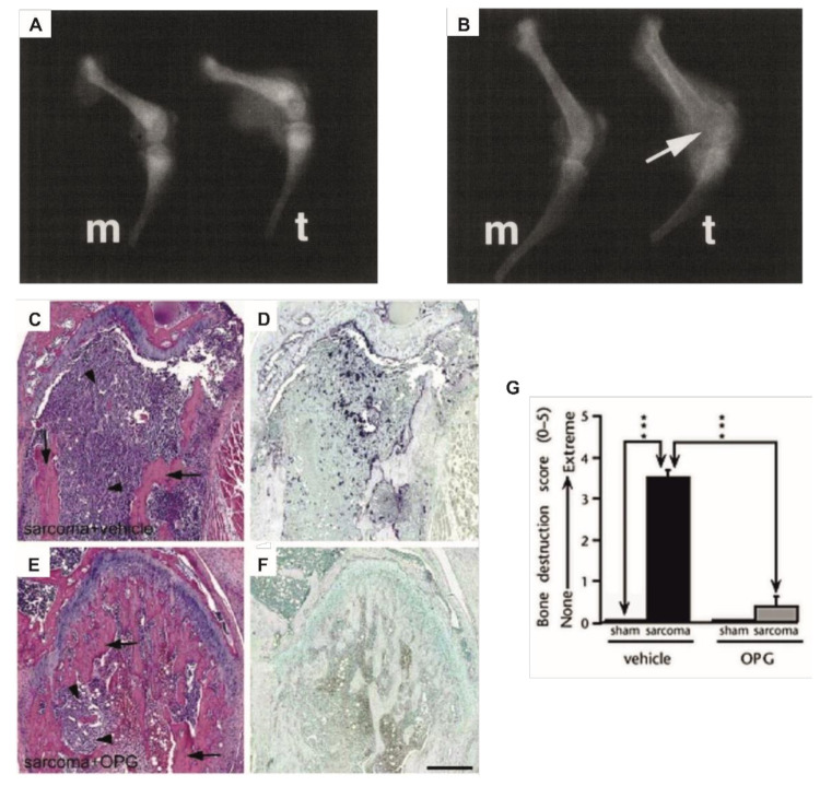
Figure 1.
Examples of a central role of osteoclasts in primary bone cancer-induced osteolysis. (A,B) X-ray images of femurs from (A) osteoclast-deficient strain B6C3Fe-a/a-Mitfmi or (B) wild type mice, 14 days after inoculation with medium alone (m) or osteolytic sarcoma cells (t). Osteolysis (arrow) was only observed in the wild type mice. (C–F) Sections of sarcoma-inoculated mouse femurs stained with hematoxylin and eosin (C,E) or tartrate-resistant acid phosphatase, staining osteoclasts dark violet (D,F). Arrows indicate bone and arrow heads indicate tumor cells. OPG treated mice (E,F) showed greatly reduced bone destruction and diminished osteoclast numbers compared to vehicle treated mice (C,D). (G) Bone destruction score in sham and sarcoma inoculated mice from the same experiment as C–F, treated with OPG or vehicle. Data are shown as mean (± s.e.m.). *** p < 0.001, one-way ANOVA, Fisher PLDS, arrows indicate the groups being compared. (A,B) Reproduced with kind permission from Clohisy and Ramnaraine, Journal of Orthopaedic Research; published by John Wiley and Sons, 1998 [63]. (C–G) Reproduced with kind permission from Honore et al., Nature Medicine; published by Springer Nature, 2000 [64].

