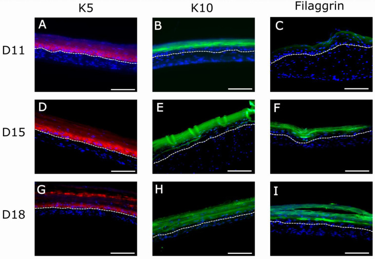
Figure 9.
Immunofluorescence (IF) analysis of organotypic skin cultures (1.2 mg/mL) using epidermis-specific markers: keratin 5 (K5), characteristic of basal epidermal cells (red staining in (A,D,G)); keratin 10 (K10), characteristic of suprabasal epidermal cells (green staining in (B,E,H)); and filaggrin, characteristic of the stratum granulosum, the last compartment containing living cells before dead and cornified cells (green staining in (C,F,I)) at three timepoints of differentiation at the air–liquid interface (11, 15, and 18 days). Blue spots in (A–I) correspond to cell nuclei stained with DAPI. The bright fluorescent lines in (E) are an artifact caused by folds present in the sample during histological processing. Dotted white line indicates the dermo-epidermal junction (basal membrane). Scale bar: 200 µm.

