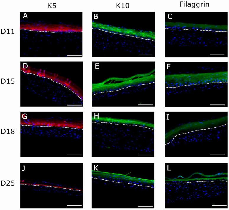
Figure 10.
Immunofluorescence (IF) analysis of organotypic skin cultures (2.4 mg/mL) of human-specific epithelial markers: K5 (A,D,G,J), K10 (B,E,H,K), and Filaggrin (C,F,I,L) at four different timepoints of development (11, 15, 18, and 25 days). Dotted white line indicates the dermo-epidermal junction (basal membrane). The bright fluorescent lines in (D) are an artifact caused by folds present in the sample during histological processing. Scale bar: 200 µm.

