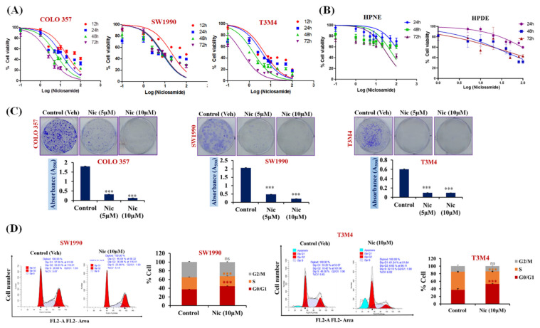
Figure 1.
Nic-mediated inhibition of cell growth, colony formation, and G1-phase cell accumulation in pancreatic cancer (PC) cells. (A,B) Concentration (100 nM–100 µM) and time-dependent (12, 24, 48, and 72 h) effect of Nic treatment on the viability of PC cells (A) and normal (human pancreatic nestin expressing (HPNE), pancreatic ductal (HPDE)) pancreatic cell lines (B) were determined by MTT assay. Values are expressed as mean ± SEM (n = 5). (C) Evaluation of in vitro colony formation capabilities of PC cell lines (COLO 357, SW1990, and T3M4) treated with Nic ~IC20 (5 μM); ~IC50 (10 μM) (upper panel). Cell colony was then dissolved in 10% glacial acetic acid, and the optical density was measured at 590 nm under a microplate reader. Quantitative analysis of these colonies represented (lower panel) and values are expressed as mean ± standard error of the mean (SEM, n = 5), p values: *** p < 0.001 vs. control, Student’s t-test. (D) SW1990 and T3M4 cells-treated with Nic (10 μM) and stained with propidium iodide (PI) followed by flow cytometry to determine the cell cycle distribution based on DNA content (left panel). Quantitative analysis of these micrographs was shown as mean ± SEM, p values: *** p < 0.001 vs. control (right panel).

