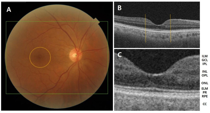
Figure 1.
(A) Anatomy of the fundus and macula (circle) in a normal eye. (B,C) Layer-by-layer B-scan swept-source optical coherence tomography display of normal retina. CC, choriocapillaris; ELM, external limiting membrane; GCL, ganglion cell layer; ILM, inner limiting membrane; INL, inner nuclear layer; IPL, inner plexiform layer; NFL, nerve fiber layer; ONL, outer nuclear layer; OPL, outer plexiform layer; PR, photoreceptor; RPE, retinal pigment epithelium.

