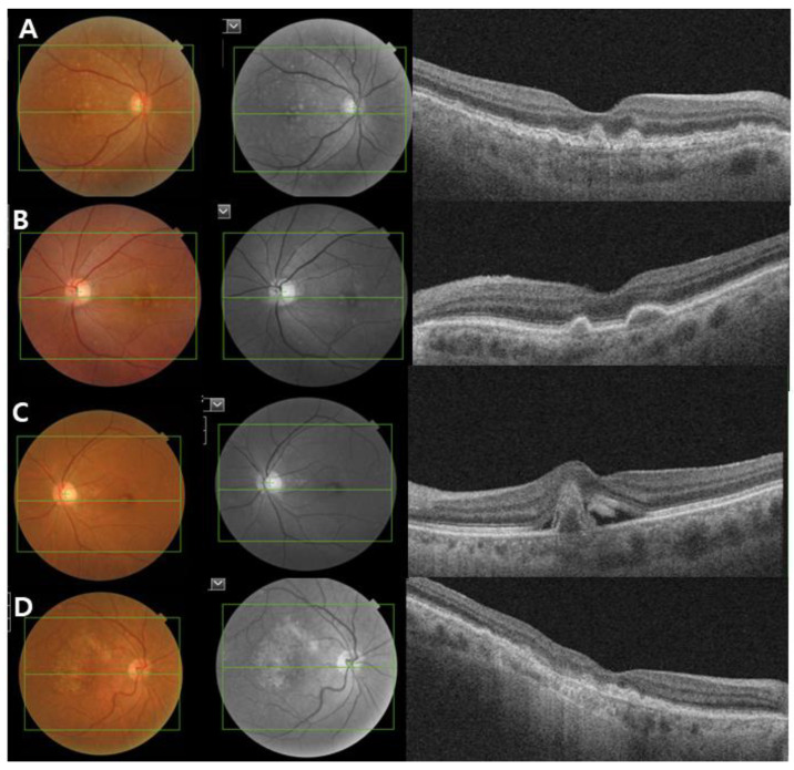
Figure 2.
Clinical image of age-related macular degeneration (AMD). Color fundus photograph, red-free fundus photograph and swept-source optical coherence tomography (SS-OCT) images showing the characteristics of early and intermediate AMD (A,B), neovascular AMD (C) and geographic atrophy (D). (A,B) Non-neovascular AMD: Images showing small and intermediate soft drusen. (C) Neovascular AMD: subretinal fluid with subfoveal hemorrhage and a large pigment epithelial detachment. (D) Geographic atrophy: a well-demarcated area of fovea-involving retinal pigment epithelium atrophy.

