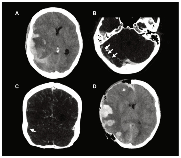
Figure 1.
(A) Preoperative CT scan which demonstrates parenchymal stasis hemorrhage and a spontaneous acute subdural hematoma with associated midline shift and impending herniation. (B,C) Axial and coronal images, respectively, which demonstrate marked sinus vein thrombosis (arrows) on CT venography (CTV). (D) Postoperative CT scan after decompressive craniectomy.

