Figure 1.
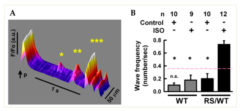
Spontaneous Ca2+ release in adult cardiomyocytes isolated from CPVT mouse hearts. (A) Fluorescence image of Ca2+ dynamics monitored with confocal microscopy in a heterozygous RyR2RS/wt (RS/WT) CM during isoproterenol (ISO, 1 μM) stimulation. Upon steady-state pacing, the last depolarization pulse (p) activates a regular intracellular Ca2+ transient, followed by abnormal spontaneous (un-paced) Ca2+ waves: * localized; ** cell-wide; *** regenerative. F/Fo, normalized fluorescence Ca2+ signal intensity. (B) Bar graph summarizing cell-wide Ca2+ wave occurrence in WT and CPVT CMs in basal conditions vs. ISO stimulation. Error bars represent SD; * p < 0.05.

