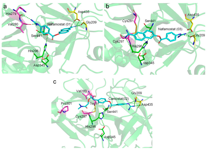
Figure 5.
Putative binding modes of Nafamostat (a,b) and Camostat (c). Ligands, catalytic triad residues, binding residues in the S1 pocket, and other important binding residues outside of the S1 pocket are shown in cyan, green, yellow, and magenta sticks, respectively. Salt bridges and hydrogen bonds are shown in green dashed lines. Poses 1 and 3 of Nafamostat represent the “forward” and “reverse” binding modes, respectively.

