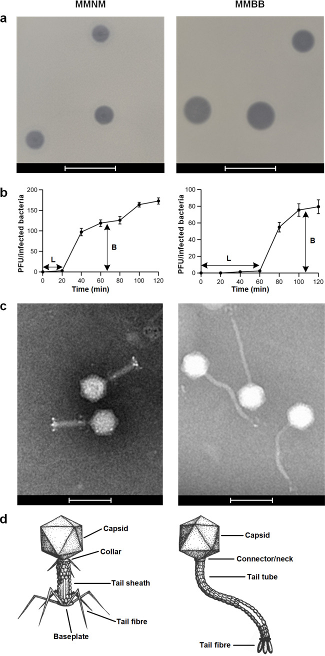
FIG 5.
Morphological characterization of phages MMNM and MMBB. (a) Plaque morphology analysis was performed using the double overlay method. Plaque morphology analysis was performed using the double overlay method after liquid infections of B5055 ΔompK36 with serially diluted MMNM and MMBB. Plaque morphologies of MMNM and MMBB were determined after overnight incubation at 37°C. Bars, 10 mm. (b) One-step growth curve of MMNM (left) and MMBB (right) was performed by coincubation with the host strain for 10 min at 37°C for phage adsorption, after which the mixture was subjected to centrifugation to remove free phage particles. The resuspended cell-phage pellets were incubated at 37°C and sampled at 10-min intervals for 120 min. L, latent period; B, burst size. Data points are the means of three biologically independent samples, and the error bars are the standard deviations. (c) Transmission electron micrographs of MMNM (left) and MMBB (right). Bars, 100 nm. (d) Based on electron microscopy (EM) micrographs, illustrations of MMNM (left) and MMBB (right) show the cognate features in Myoviridae and Siphoviridae with annotation.

