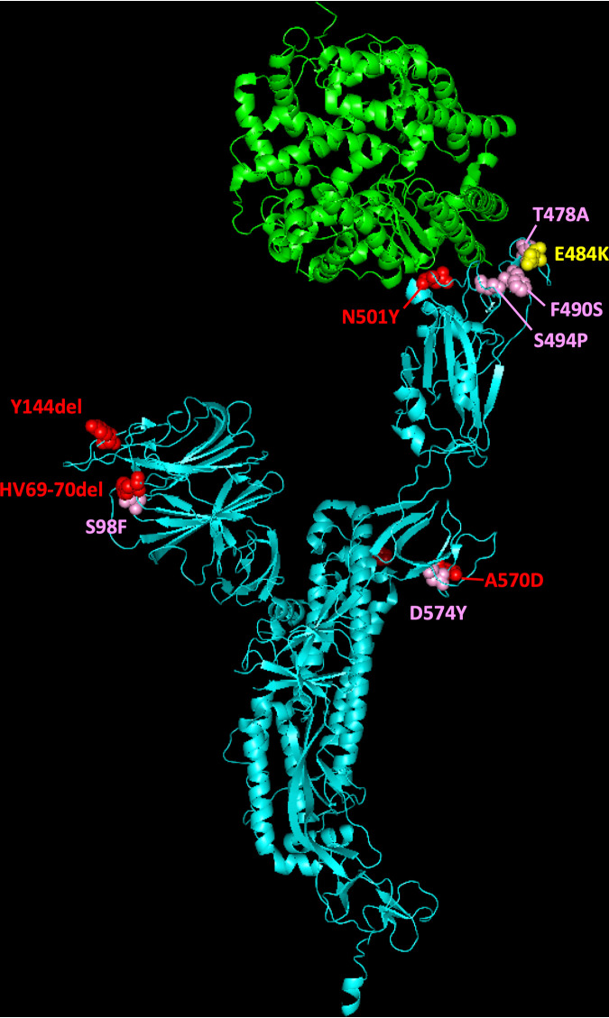
FIG 5.
Molecular cartoon diagram of the 3D structure of SARS-CoV-2 spike protein monomer in the open form (cyan) in complex with receptor ACE-2 (green). The image was generated using PyMOL Molecular Graphics System version 1.7.0.3 software (Schrödinger, LLC) using cryo-EM data (Protein Data Bank accession number 7DF4 [36]). The location of selected amino acid substitutions found in SARS-CoV-2 B.1.1.7 variant are shown in red; mutations found at low frequency in viral RNAs from sewage are indicated in pink, and the position of mutation E484K present in B.1.351 and P.1 VOCs is shown in yellow.

