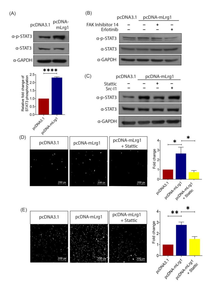
Figure 5.
Lrg1-induced activation of the EGFR/STAT3 pathway is required for melanoma cell invasiveness. (A) Representative Western blot (left) and densitometry (right) analyses (right) showing the levels of phospho-STAT3 (Tyr705) and total STAT3 in Lrg1 overexpressing B16F10 cells. GAPDH was used as a loading control. (B) Representative images of Western blot analysis showing the levels of phospho-STAT3 (Tyr705), STAT3, and GAPDH in Lrg1 overexpressing B16F10 cells subjected to treatment with FAK inhibitor 14 (FAK inhibitor) or erlotinib (EGFR inhibitor). (C) Representative images of Western blot analysis showing the levels of phospho-STAT3 (Tyr705) and total STAT3 in Lrg1 overexpressing B16F10 cells subjected to treatment with Src-I1 (Src inhibitor) and stattic (Stat3 inhibitor). GAPDH was used as a loading control. (D) Representative images and quantification of migrated Lrg1 overexpressing B16F10 cells subjected to stattic treatment. (E) Representative images and quantitative analysis of invaded Lrg1 overexpressing B16F10 cells subjected to stattic treatment. All images are representative. Data are presented as mean ± SEM of three independent experiments. Statistical analyses were performed via two-tailed, unpaired Student’s t-test. * p < 0.05; ** p < 0.01; **** p < 0.0001.

