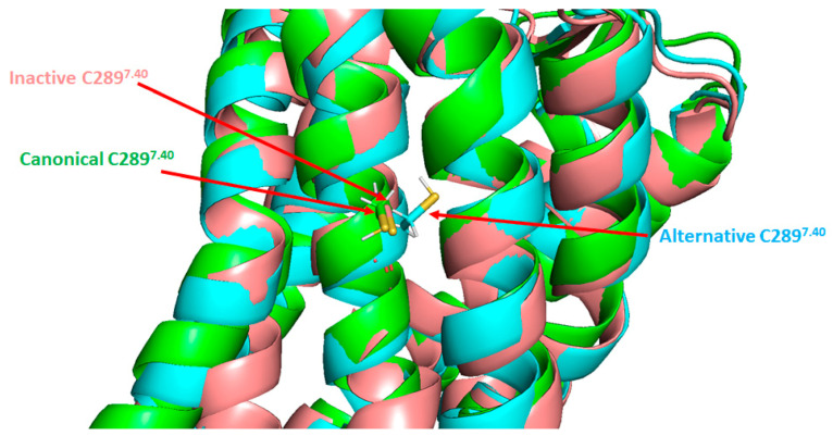
Figure 7.
Positions of the cysteine 289 residue in the three known AT1 structures. Superimposition of the three known AT1 structures: inactive (salmon), canonical active (green) and alternative active (cyan). TM6 and TM7 are located in the front. C289 residue is indicated by arrows. The reference inactive structure of AT1 has been chosen as PDB code 4YAY (from Zhang et al. [99]). Canonical active and alternative active structures are simulation data available in supplementary material from Suomivuori et al. [98]. The figure was prepared with the PyMOL software (The PyMOL Molecular Graphics System, Version 2.5.0. Schrödinger, LLC).

