Figure 4.

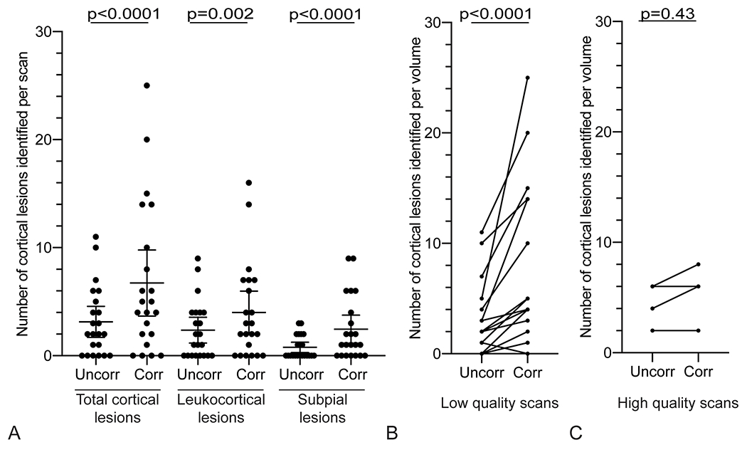
More cortical lesions are identified on navigator-guided T2*w images that have undergone motion and B0 correction. (A) More total cortical lesions, leukocortical lesions, and subpial lesions were identified on corrected images (“Corr”) compared to the respective uncorrected images (“Uncorr”). (B) The number of cortical lesions identified in cases with low-quality uncorrected images (average rating <3) increased with correction in 14/18 scans (78%), vs an increase in cortical lesion identification in 2/4 cases (50%) with high-quality uncorrected scans (average rating ⩾3) (C). Quality ratings: 1 – severe motion artifacts, 2 – moderate motion artifacts, 3 – minimal motion artifacts, 4 – no motion artifacts.
