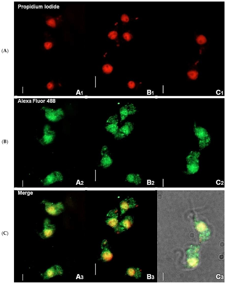
Figure 1.
Immunofluorescence staining of T. brucei bloodstream form suggests cytosolic localization of TbHsp70.c. Distribution of TbHsp70.c in T. b. brucei cells using TbHsp70.c primary antibody, and Alexa Fluor 488 secondary antibody (A–C). Parasite DNA was stained with propidium iodide. Upright scale bar, 5 µm. Rows: Propidium iodide—parasite DNA, including the nucleus and kinetoplast detected with a UV filter, shown in red; Alexa Fluor 488—localization of TbHsp70.c; Merge—amalgamated image of DNA staining and TbHsp70.c localization.

