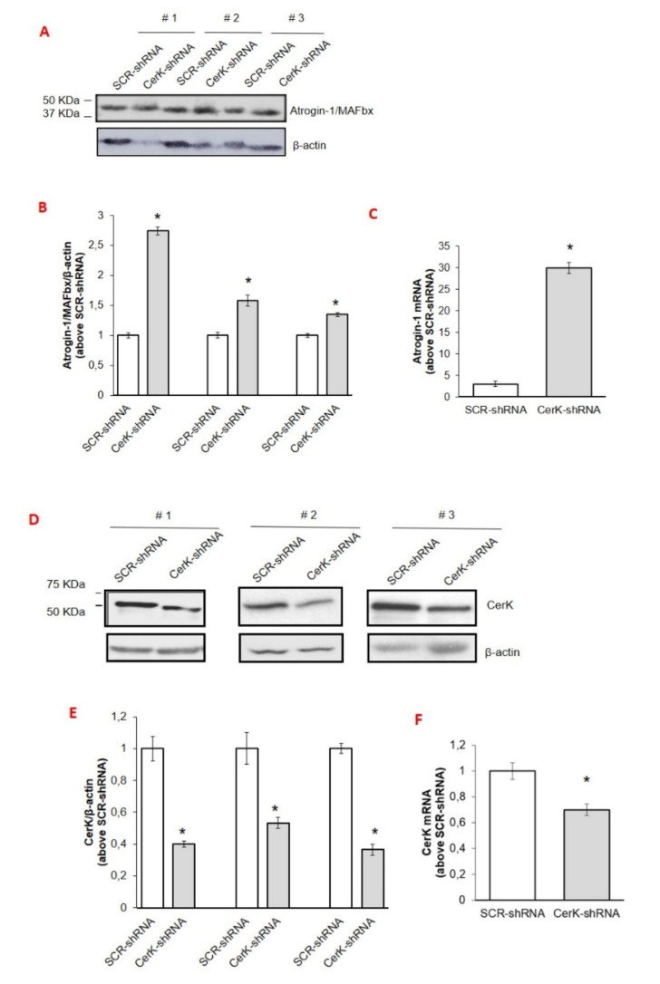
Figure 4.
Effects of CerK silencing on atrogin-1/MAFbx and CerK protein and mRNA expression in tibialis muscle tissues (A–F). Cell lysates for atrogin-1/MAFbx (A) and CerK (D) and protein expression and total RNA for atrogin-1/MAFbx mRNA expression (C) or CerK (F) were obtained from tibialis muscle tissues of healthy mice electroporated with scrambled shRNA (SCR-shRNA) or shRNA specific to CerK (CerK-shRNA) as described in Methods. Healthy mice were randomized into groups, SCR-shRNA (n = 3) and CerK-shRNA (n = 3). Tissue lysate proteins (40 µg), obtained from tibialis muscle tissues of healthy mice transfected with SCR-shRNA or CerK-shRNA, were subjected to SDS-PAGE and immunoblotted as reported in Methods. A blot representative of at least three independent experiments and densitometric analysis is shown (B,E). Normalized data (means ± SEM) to the β-actin bands, are reported in the graph (Student’s t test, * p < 0.05 vs. SCR-shRNA). (C,F): Total RNA was prepared and reverse transcripted and Real-time PCR performed using primers for atrogin-1/MAFbx (C) or CerK (F) as reported in Methods. Data are presented as fold change (mean ± SEM) of at least 3 independent experiments (Student’s t test, * p < 0.05 vs. SCR-shRNA).

