FIGURE 4.

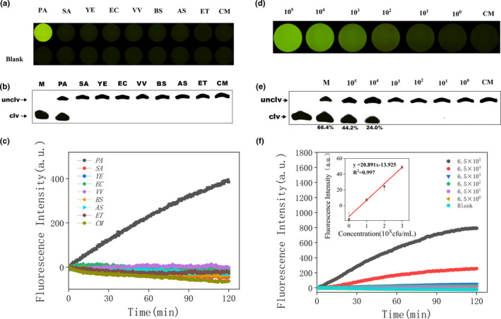
(a) Iindicator plate images of PAE‐1 specific detection after 10 min. PA: Pseudomonas aeruginosa; SA: Staphylococcus aureus; YE: Yersinia enterocolitica; EC: Escherichia coli; VV: Vibrio vulnificus; BS: Bacillus subtilis; AS: Aeromonas salmonicida; ET: Edwardsiella tarda; CM: Blank complete medium. (b) Image of cleavage gel for PAE‐1 specificity detection within 2 hr. (c) Biosensor‐based assay of PAE‐1 in blank culture medium and seven different bacteria. Gel image (e), indicator plate (d), and biosensor measurement (f) of PAE‐1 in different concentrations of Pseudomonas aeruginosa in CEM‐PA. (f) Fluorescence values of P. aeruginosa at different concentrations (Blank: ddwater without P. aeruginosa). Inset: Analytical calibration curve of fluorescence values of P. aeruginosa (concentrations of 6.5, 6.5 × 10, 6.5 × 10², 6.5 × 10³) for 2 hr
