
Figure 1.
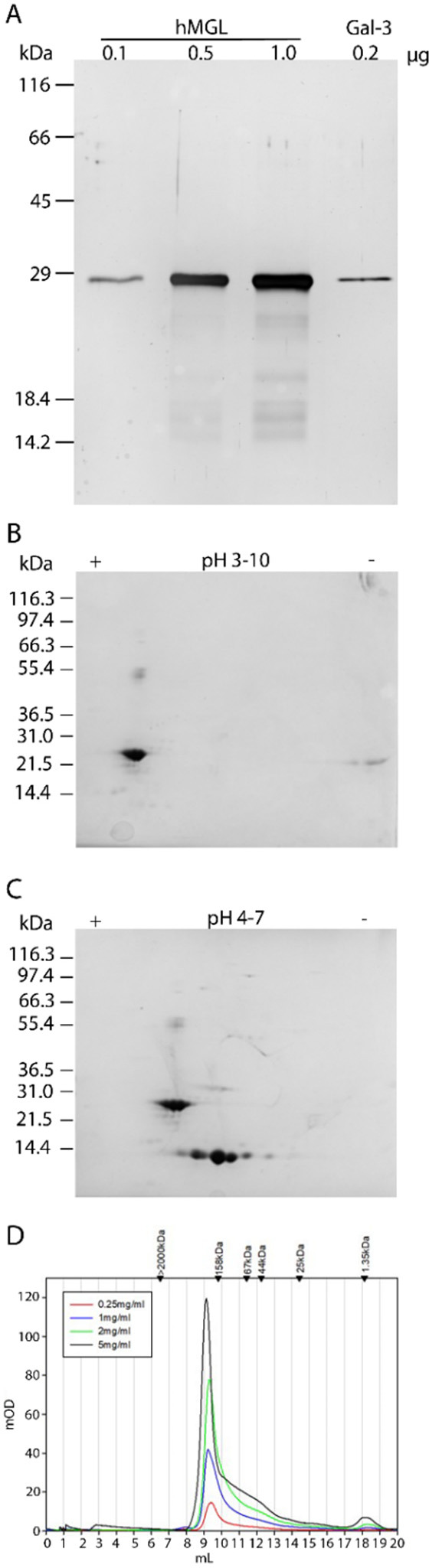
Characterization of hMGL by SDS–PAGE, two-dimensional (2D) gel electrophoresis, and gel filtration. (A) SDS–PAGE (15% polyacrylamide) of 0.1, 0.5, and 1.0 μg of hMGL as well as 0.2 μg of galectin-3 (protein with a similar mass and constitution with stalk and CRD). (B) 2D gel electrophoresis of 20 μg of hMGL in the pI range of 3–10. (C) 2D electrophoresis of a mixture of 20 μg of hMGL and 20 μg of galectin-1 in the pI range of 4–7. (D) Gel filtration of hMGL at the indicated concentrations (applied volume of 50 μL in each case).