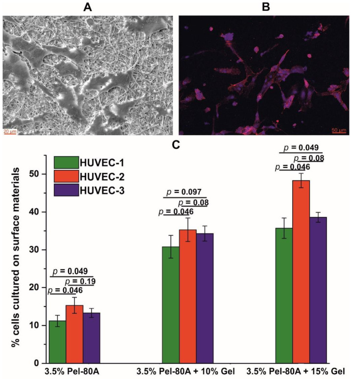
Figure 2.
Interaction of endothelial cells with electrospun Pel-80A-based scaffolds. SEM image of endothelial cells cultured on 3.5% Pel-80A + 10% Gel scaffold (A). Fluorescence image of cell morphology of HUVEC cultured on the surface of the scaffold containing 3.5% Pel-80A + 15% Gel (nuclei and actin filaments of cells stained with Hoechst 33342 and Phalloidin-TRITC dye, respectively) (B). Viability of HUVEC from 3 different donors on the surface of different 3D matrices after 48 h of cultivation (data are presented as the mean of three replicates with standard deviation) (C).

