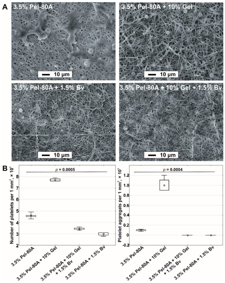
Figure 3.
Blood contact test for different scaffolds. SEM images obtained after incubation of electrospun tubes with blood (scale bar, 10 μm; magnification, 1000×) (A). Quantitative data of adhered platelets and their aggregates on surface tubes are presented as a median and interquartile range (B).

