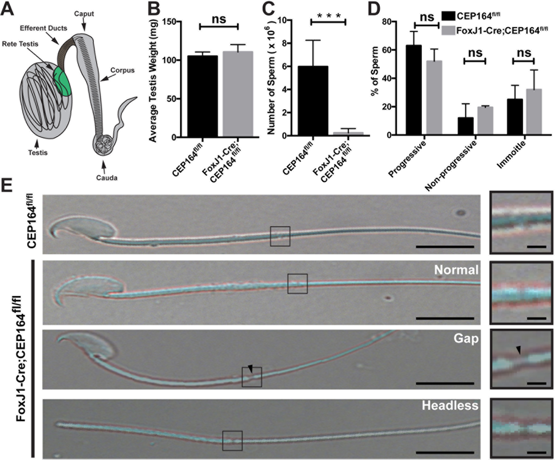
Fig 1. Loss of CEP164 leads to reduced sperm counts.

(A) Schematic of the male reproductive system. (B) Testes were isolated from 2 to 4-month-old CEP164fl/fl and FoxJ1-Cre;CEP164fl/fl mice and weighed (N=5 per genotype). Error bars represent means ± SD. ns, not significant. (C) Sperm were collected from the cauda epididymis of 2 to 4-month-old mice and counted on a hemocytometer (N=5 per genotype). Error bars represent means ± SD. ***, p<0.001. (D) Sperm motility. A total of 100 sperm from 3 mice per genotype were scored as either progressive (linear movement), non-progressive (circular movement), or immotile (no movement). Error bars represent means ± SD. ns, not significant. (E) Representative images of isolated sperm. The boxed regions are magnified on the right. Arrowheads point to a gap between the midpiece and the principal piece. Scale bars, 5 µm and 1 µm (magnified images).
