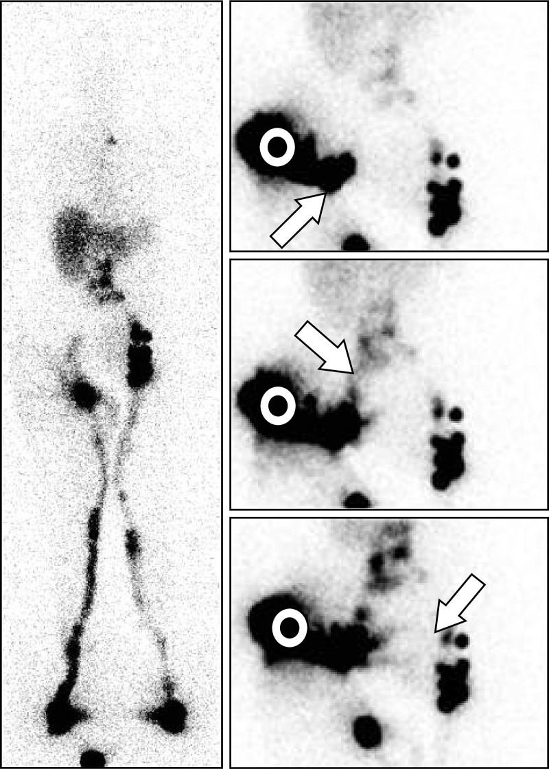
Fig 7. “Intraabdominal LNs and collateral vessels are shown in a case of secondary LLLE!”.
- On the left side, one normal draining LV reaching one complete infradiaphragmatic LN axis was observed.
- On the right side, one draining LV reaching one inferior inguinal LN from which lymphatic reflux was observed with one area of faint activity in the upper inguinal area but no common iliac LNs.
The three consecutive images [right: From top to bottom, anterior views] centered on the pelvis and abdomen [white circles] obtained after the intradermal injection performed on the same day in the external part of the buttock revealed draining LVs toward the inguinocrural LN [from bottom to top and left to right, oblique arrow], the common iliac LNs [from up to down and left to right, arrow] and toward the contralateral inguinal LNs in the prepubic area [from top to bottom and right to left, arrow]. Manual lymphatic drainage applied to the root of the right limb allowed reduction of the lymphedema from + 20.6% excess to + 4.9% excess [determined by comparison of the sum of the circumference values measured every 4 cm from the ankle to the root of the limb].

