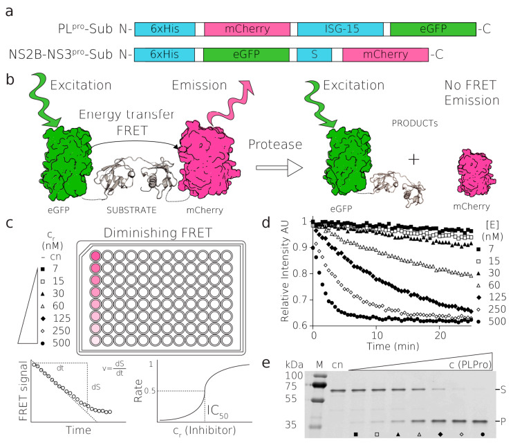
Figure 1.
Substrates, principles of FRET assay. (a) The fluorescent PLpro and NS2B-NS3pro substrates, (b) schematics of the FRET-based proteolytic assay, eGFP is excited with a green light at 488 nm, FRET transfers the excitation to mCherry which emits the light that is detected. After proteolytic cleavage, this FRET signal is abolished, (c,d) the reaction of PLpro substrate with a serial dilution of PLpro enzyme (500–7 nM), (c) schematics of reactions followed in the fluorescent plate reader, (d) these reactions were loaded on SDS-PAGE gel and visualized on a fluorescent scanner (e), where (S) and (P) donate substrate and product bands and full-length gel are shown in Figure S8.

