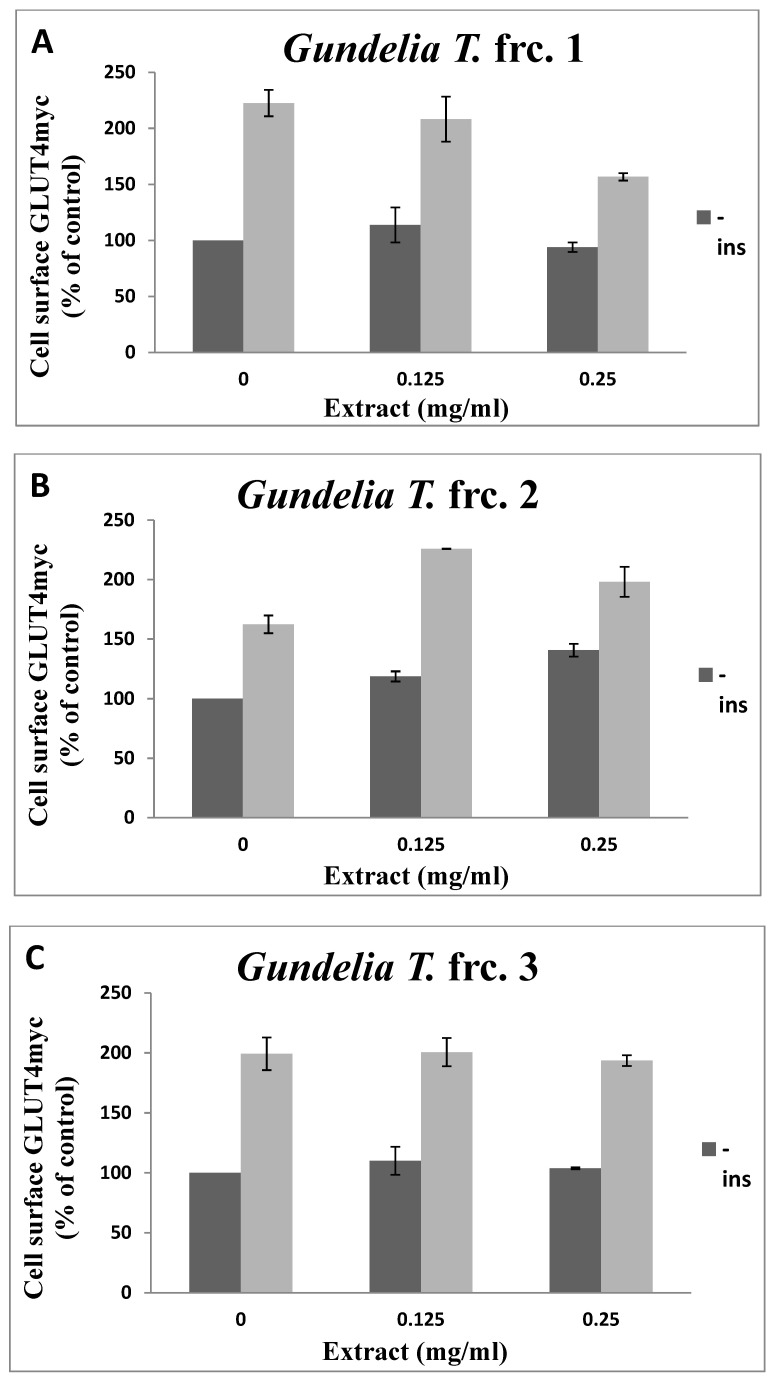
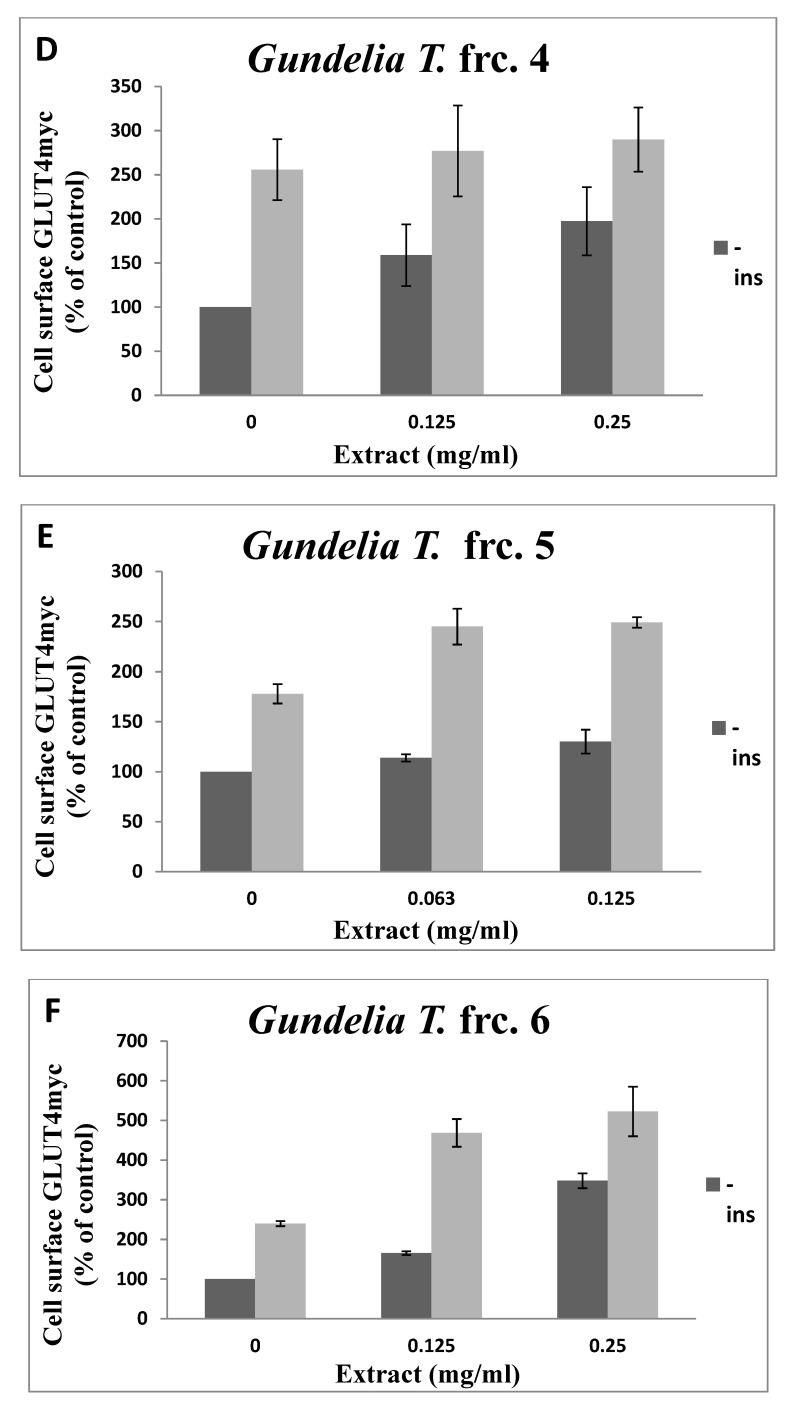
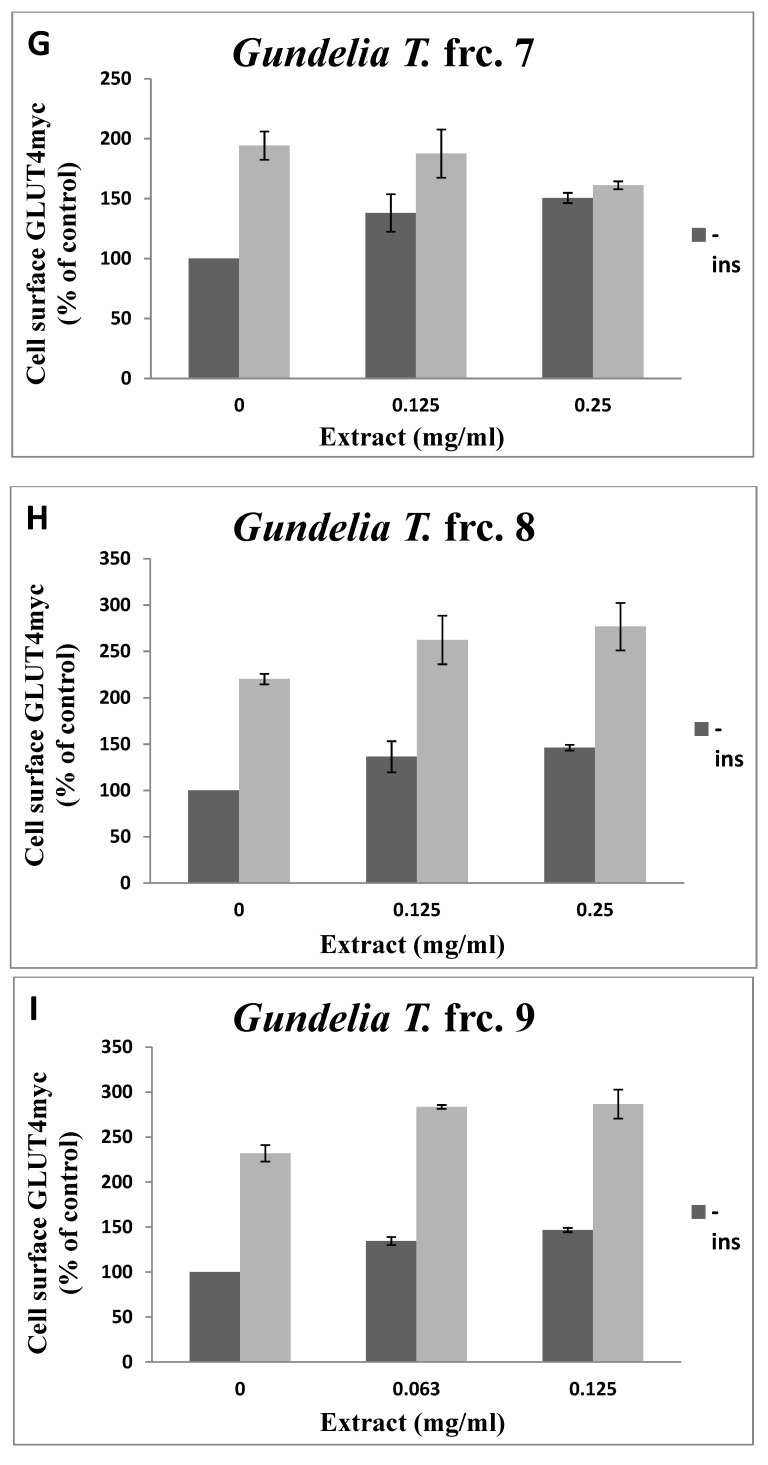
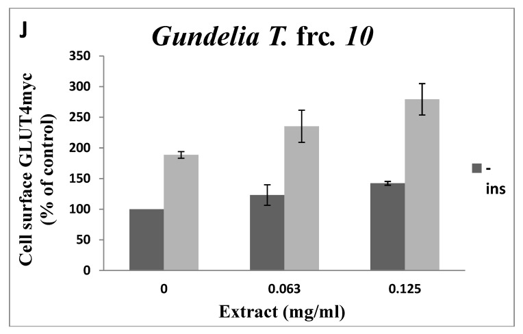
Figure 3.
GLUT4 translocation to the plasma membrane. For the evaluation of the GLUT4 L6-GLUT4myc, cells (150,000 cell/well) were exposed to GT fractions (A–J) for 20 h. Serum depleted cells were treated without (−) or with (+)1 µM insulin for 20 min at 37 °C and surface myc-tagged GLUT4 density was quantified using the antibody coupled colorimetric assay. Values given represent means ± SEM (relative to untreated control cells) of three independent experiments carried out in triplicates.


