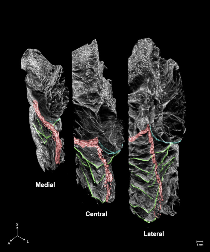
Fig 1. Micro-computed tomography (microCT) images of the orbicularis retaining ligament (ORL).
The number, complexity, and ambiguity of the fibers comprising the ORL increased anteriorly and laterally. Green, blue, and red indicate the ORL, the orbital septum and, the section of the orbicularis oculi. S, superior; A, anterior; L, lateral.

