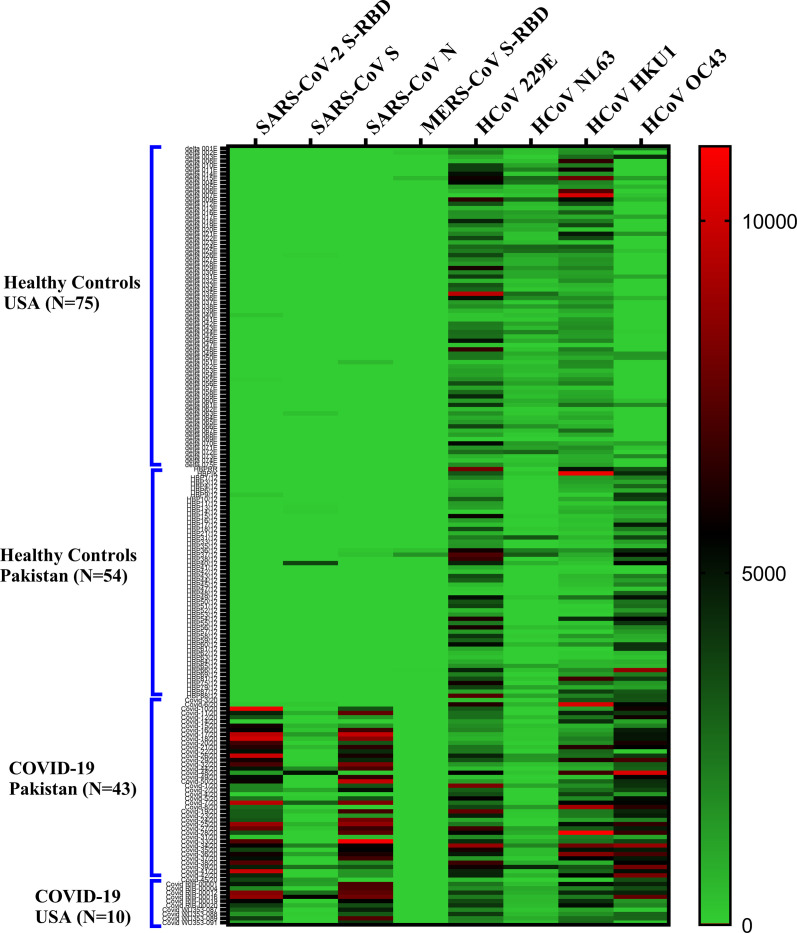
Fig 2. Heat map depicting overall antibody responses detected by multiplex microbead panel against members of the coronavirus family (SARS-CoV-2; SARS-CoV; MERS-CoV and 4 common coronaviruses (229E, NL63, OC43, HKU1)) in COVID-19 patients and healthy controls.
Antibody responses (IgG) to SARS-CoV-2 S-RBD; SARS-CoV S and N; MERS-CoV S-RBD; and S proteins of 4 common coronaviruses are shown. In patients where multiple time points were available, the latest time point after the onset of symptoms is shown. Each row corresponds to one sample and columns correspond to CoV antigens in the multiplex assay. The color intensity scale represents the relative MFI (median fluorescent intensity) values ranging from the highest (10,000; red) to no antibody response (0; green). No significant background reactivity to SARS-CoV-2 S-RBD, SARS-CoV S, SARS-CoV N, and MER S-RBD was detected in healthy controls from US and Pakistan.

