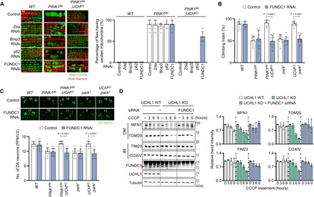
Fig. 3. Mitophagy induced by UCHL1 deficiency is mediated by FUNDC1.

(A) Left: Confocal fluorescence images of the adult flight muscles. Right: Percentage of the flies having swollen mitochondria. n = 10. Green denotes mitochondria, and red denotes actin filament. Scale bar, 5 μm. (B) Measurement of the climbing ability in the adult flies. n = 10. (C) Confocal immunofluorescence images and numbers of DA neurons in the PPM1/2 regions of adult fly brains. n = 10. Green denotes DA neuron. Scale bar, 20 μm. The number in panels indicates the number of DA neurons in each image. (D) Left: Immunoblot analysis of mitochondrial outer (MFN1 and TOM20) and inner (TIM23 and COXIV) membrane proteins upon 20 μM CCCP and FUNDC1 small interfering RNA (siRNA) treatment. Right: Relative quantification of the immunoblot band intensity of indicated mitochondrial proteins. n = 3. Two-way ANOVA with Tukey’s multiple comparison test was used (B and C). All data were presented as means + SD.
