
Fig. 2. Shc associates with Alk5 and promotes shear stress–induced EndMT.
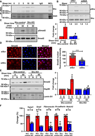
(A) Coimmunoprecipitation of Alk5 with Shc after shear stress. n = 3. IgG, immunoglobulin G. WCL, whole cell lysate. (B) Scr or Alk5 siRNA–transfected ECs were exposed to chronic oscillatory shear stress; Shc was immunoprecipitated and its Y239,240 phosphorylation was assayed. n = 3. (C) ECs from Shc-Control and Shc-KO mice were exposed to shear stress, and phosphorylation of Smad2 was determined. (D) Scr or Shc siRNA–transfected BAECs were exposed to shear stress; nuclear translocation of Smad2 was determined by immunofluorescence staining and quantified as mean fluorescence intensity on ImageJ. (E) ECs from Shc-control and Shc-KO mice were exposed to shear stress and separated into cytosolic and nuclear fractions. Western blots, performed using antibodies against the cytoplasmic protein Hsp90 and nuclear protein Lamin B1, indicated that the extracts are virtually free from cross-contamination. Hsp90 served as the loading control for cytoplasmic extracts and Lamin B1 for nuclear extracts. (F) ECs from Shc-Control and Shc-KO mice were exposed to chronic shear stress; qPCR was performed to quantify the expression of the EndMT markers Twist1, Snail, fibronectin, N-cadherin, and Notch3. Scale bar, 20 μm; n = 4. Data are presented as means ± SEM. P values were obtained by two-tailed Student’s t tests using GraphPad Prism. *P < 0.05; **P < 0.01, ****P < 0.001, and #P < 0.05 relative to the respective shear time point of Scr siRNA.