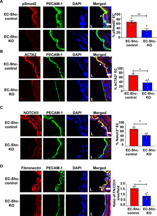
Fig. 3. Shc mediates EndMT in disturbed shear stress regions in vivo.

(A to D) Sections of the LCA from mice that underwent partial carotid ligation in the LCA, followed by 3 weeks of high-fat diet feeding, were immunostained with the phosphorylated form of the EndMT intermediary Smad2 and EndMT markers ACTA2, Notch3, and fibronectin. Staining was also performed with PECAM-1 and DAPI to identify the endothelium and nuclei, respectively. Positive cells (defined as coexpressing PECAM-1 and the respective EndMT marker) were quantified and shown by arrowheads. Scale bars, 10 μm; n = 4 EC-Shc-control mice and 4 EC-Shc-KO mice. Data are presented as means ± SEM. P values were obtained using two-tailed Student’s t tests using GraphPad Prism. *P < 0.05; **P < 0.01.
