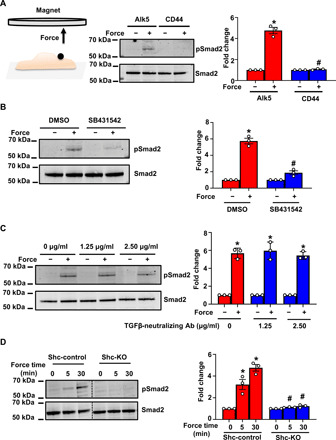
Fig. 5. Force on Alk5 specifically induces mechano-EndMT via Shc.

(A) Mouse ECs were incubated with anti-Alk5 or CD44 (negative control) antibody–coated beads and subjected to force (10 pN). Phosphorylation of Smad2 was determined by Western blotting and quantified using Image Studio Lite v.5.2. n = 3 (B and C) Mouse ECs were treated with the (B) Alk5 kinase inhibitor SB431542 or (C) varying concentrations of a TGFβ-neutralizing antibody (Ab), incubated with anti-Alk5–coated beads and subjected to force before analysis of the phosphorylation of Smad2. n = 3. Data are presented as means ± SEM. P values were obtained using two-tailed Student’s t test using GraphPad Prism. DMSO, dimethyl sulfoxide. (D) Shc-control and Shc-KO ECs were incubated with anti-Alk5–coated beads and subjected to force application before analysis of the phosphorylation of Smad2. n = 3. Data are presented as means ± SEM. P values were obtained using two-tailed Student’s t test using GraphPad Prism. Phosphorylated proteins are indicated by “p-.” *P < 0.05 relative to the no-force condition, and #P < 0.05 relative to the respective force application time point with Alk5.
