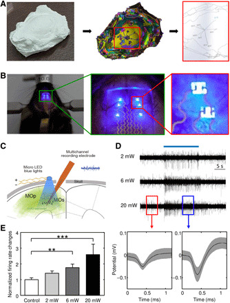
Fig. 6. Scalp-mounted optogenetic stimulator with topography-based dry transfer printing.

(A) 3D scanning process of the mouse brain model for obtaining the spatial information of stimulus sites considering the biological anatomy. Photo credit: Seungkyoung Heo, Daegu Gyeongbuk Institute of Science and Technology. (B) Optical images of an optogenetic stimulator resting on the target positions of the mouse brain during the operation of the μ-ILEDs and magnified optical images of a device with active components and stretchable interconnections. Photo credit: Seungkyoung Heo, Daegu Gyeongbuk Institute of Science and Technology. (C) Schematic diagram for the in vivo experiment. The optogenetic stimulator sheds blue light on the motor cortex, activating ChR2. Inserted multichannel electrodes simultaneously record changes in neural activity in response to optogenetic stimulation with various intensities. (D) Example traces of neural activity with blue light illumination (top) and examples of spike waveforms of isolated units evoked by the μ-ILED operation (bottom) (ON, red box; OFF, blue box). (E) Frequency of the firing rate upon optogenetic stimulation at various intensities (0, 2, 6, and 20 mW).
