FIGURE 2.

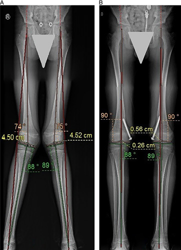
Bilateral genu valgus; femoral origin. A, Long-standing radiograph scanogram of a skeletally immature patient. B, The same patient after using the retrograde transphyseal guided growth technique with optimal bilateral deformity correction.

Bilateral genu valgus; femoral origin. A, Long-standing radiograph scanogram of a skeletally immature patient. B, The same patient after using the retrograde transphyseal guided growth technique with optimal bilateral deformity correction.