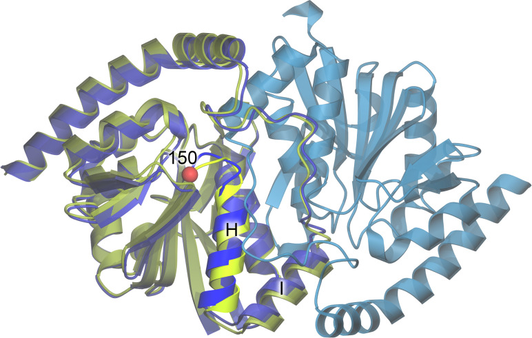
FIG. 3.
Structure of ICH. The ribbon diagram for the WT ICH dimer is shown in blue, with protomer A colored darker blue and protomer B lighter blue. The structure of G150T ICH (yellow-green) is superimposed on protomer A of WT ICH. The location of residue 150 is represented as a red sphere, and the mobile helix is labeled H and shown in brighter colors.

